Download our anal examination diagnostic flowchart for a step-by-step guide on approaching a patient who needs an anal examination.
Anorectal examination
Indications
- Assess symptoms such as itch, pain, bleeding, discharge, lumps, or change in bowel habit
- Examine the prostate
- Screen for anal cancer screening in high risk patients (such as MSM)
Preparation
Equipment
- Examination table
- Good lighting
- Gloves
- Lubricant
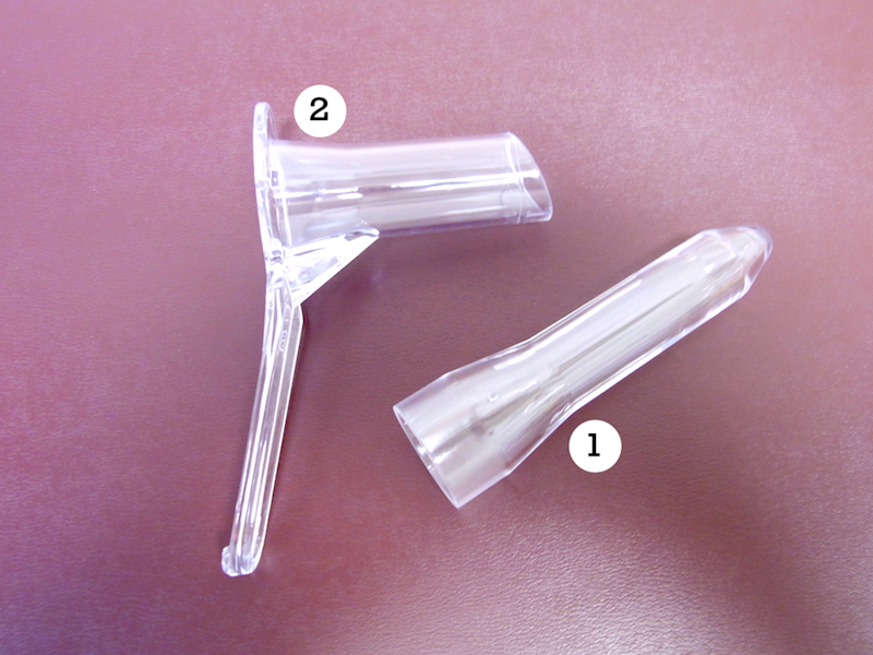
- Anoscope if required (also called a proctoscope)
- Swabs (if required)
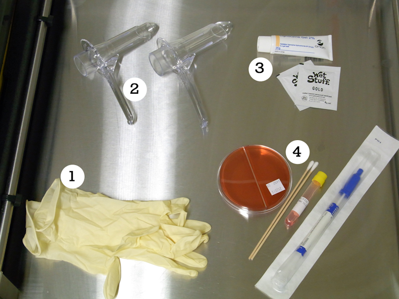

Anorectal examination equipment
Positioning
- In lateral decubitus position with knees bent towards the chest
- It may be helpful to ask the patient to lift the right gluteal area with the right hand to expose the perianal area

Warning
May contain graphic images of human anatomy, medical conditions and medical procedures. Viewing discretion is advised.
Anorectal examination positioning
Inspection
Visual inspection
By parting the perianal skin, visual inspection can reveal:
- fissures
- fistulae
- perianal dermatitis
- lumps such as haemorrhoids and warts
- lesions such as ulcers of herpes or syphilis
Palpation
Digital anal examination (DARE) should always be done unless the patient is experiencing severe pain (such as fissure or abscess).
Insert one lubricated finger and sweep 360 degrees around the entire anal canal. The prostate should be identified, as well as any palpable masses or nodules. Columns of Morgagni may be recognised.
How to perform a digital anal examination
This video shows you how to perform a digital anal examination.
Anoscopic examination
Examine the perianal area for any lesions which preclude anoscopy, such as traumatic anal fissure.
Explain the examination process to the patient, reassuring them that they will feel pressure but no pain.
Complete the examination:
- Generously lubricate the anoscope with standard lubricating jelly or xylocaine jelly.
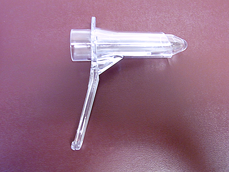
- Ensure the introducer is at the furthest point within the anoscope to avoid skin pinching.
- Insert the anoscope gently and advance it slowly in the direction of the umbilicus. If resistance due to contraction of the external anal sphincter occurs, constant gentle pressure on the anoscope will eventually fatigue the muscles and permit slow insertion.
- Maintain pressure over the introducer with the thumb during insertion to keep it from slipping out. To avoid pinching the anal mucosa, completely remove the anoscope and reinsert the device if the introducer slips or falls out during insertion.
- Once the anoscope is adequately inserted, remove the introducer and ensure good lighting.
- As the anoscope is slowly withdrawn, the anal mucosa can be visualised over the entire circumference of the canal. Any suspicious areas can be swabbed at this point.
- As the anoscope is withdrawn at the anal verge, spasm of the external sphincter may lead to rapid expulsion. Maintain firm counter-pressure on the instrument to prevent this.
Process any swabs taken.
How to perform an anoscopy
This video describes how to perform an anoscopy.